
- Scientific Area
- Dermatologic surgery
SCIENTIFIC AREA
Mohs Skin Micrographic Surgery
Mohs micrographic surgery is a precise, tissue-sparing, margin-controlled surgical technique for skin cancer treatment in which the surgeon removes the tumor in stages and examines microscopically 100% of the peripheral and deep margins during the procedure, repeating excision only where cancer persists.
It is considered the most accurate surgical method for many high-risk, recurrent, or anatomically critical skin tumors, particularly on the face.
Core Principle of Mohs Surgery
The key innovation of Mohs surgery is complete circumferential peripheral and deep margin assessment (CCPDMA). Unlike standard excision—which samples only a small fraction of margins (“bread-loafing”)—Mohs examines essentially the entire surgical margin.
This enables:
Maximal cure rates
Minimal removal of healthy tissue
Precise mapping of residual tumor
Immediate reconstruction once margins are clear
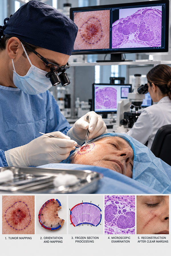
- STEP BY STEP
Classic Mohs Procedure
01
Clinical tumor definition
The dermatologic surgeon:
Examines the lesion clinically
Uses dermoscopy if useful
Defines visible margins
Sometimes marks subclinical extension
The tumor is mapped on a surgical diagram.
02
Debulking stage
The clinically visible tumor bulk is often removed first (“debulking specimen”).
The specimen may be sent for:
Permanent histopathology
Tumor subtype confirmation
Assessment of aggressive features
03
First Mohs stage
A thin saucer-shaped layer of tissue is excised around and beneath the tumor.
The excision is beveled (usually ~45°) to allow flattening of the specimen for complete margin visualization.
04
Tissue mapping and orientation
This is one of Mohs surgery’s defining features.
The specimen is:
Color-coded with dyes + Oriented anatomically + Mapped precisely
For example:
Red = superior margin
Blue = inferior margin
A detailed map links microscopic findings to exact patient anatomy.
05
Frozen section processing
The tissue is processed in-house during surgery.
The specimen is:
Frozen
Flattened
Sectioned horizontally
Mounted on slides
This differs fundamentally from standard pathology processing.
06
Microscopic examination
The Mohs surgeon examines the slides personally, to assess:
Peripheral margins
Deep margins
Tumor subtype
Perineural invasion
Residual microscopic extensions
If residual cancer remains, its exact location is marked on the map.
07
Additional stages (if needed)
Only areas containing tumor are re-excised. Instead of enlarging the whole wound, the surgeon removes targeted additional tissue.
This cycle repeats until “Tumor-free margins” are achieved.
08
Reconstruction
After oncologic clearance, the defect is reconstructed.
Options include:
Secondary intention healing
Linear closure
Skin grafts
Local flaps
Cartilage support (nose/ear)
Often reconstruction is performed the same day.
Why Mohs Differs from Standard Excision
Standard excision
The surgeon removes the tumor with predetermined margins. The specimen undergoes vertical bread-loaf sectioning, meaning only a small percentage of margins are sampled. Residual tumor can be missed.
Mohs surgery
Nearly 100% margin control is achieved intraoperatively. This gives superior cure rates while preserving normal tissue.
Main Indications of Mohs Surgery
Mohs is particularly useful for:
High-risk anatomic sites (“H-zone” of the face)
Nose
Eyelids
Lips
Ears
Temple
Periocular area
High-risk tumors
Recurrent tumors
Aggressive histology
Large tumors
Poorly defined margins
Perineural invasion
Immunosuppressed patients
Especially:
Basal-cell carcinoma
Cutaneous squamous cell carcinoma
Selected Melanoma
Rare adnexal tumors
Cure Rates
Published literature generally reports:
Primary BCC: ~98–99%
Recurrent BCC: ~94–97%
Primary SCC: ~95–99% especially in expert centers.
Variants of Mohs Micrographic Surgery
Several techniques evolved from classic Mohs to adapt to different tumors and pathology requirements.
1. Fresh tissue Mohs (modern/classic Mohs)
This is the current standard approach.
Features:
Frozen sections
Same-day margin assessment
Immediate reconstruction
Multiple stages in one visit
Most contemporary Mohs surgery uses this method.
2. Fixed tissue Mohs (chemosurgery)
The original technique described by:
Frederic E. Mohs used zinc chloride paste fixation before excision.
Process
Tissue chemically fixed in vivo
Sequential excision performed later
Advantages:
Historical control of deep tumors
Disadvantages:
Painful
Time-consuming
Tissue destruction
Largely obsolete
Today it is rarely performed.
3. Slow Mohs (paraffin section Mohs)
Also called: staged excision with permanent sections
This is especially important for tumors difficult to assess on frozen sections.
Technique: Instead of frozen tissue:
Tissue is processed in paraffin
Permanent histology used
Results obtained over 24–72 hours
Reconstruction delayed or temporary
Best indications
Especially valuable for:
Lentigo maligna / melanoma in situ
Melanocytic lesions harder to interpret on frozen sections.
Rare tumors
Dermatofibrosarcoma protuberans
Microcystic adnexal carcinoma
Sebaceous carcinoma
Extramammary Paget disease
Advantages
Superior histologic detail
Better melanocyte assessment
Immunostaining possible
Disadvantage
Delayed repair
4. Mohs with immunohistochemistry (IHC-Mohs)
Special stains are added during Mohs processing. Used for tumors difficult to identify microscopically.
Examples:
Melanoma Mohs
Markers:
MART-1
SOX10
MITF
SCC/BCC difficult cases
Cytokeratins
Rare adnexal neoplasms
EMA
BerEP4
CEA
This approach has expanded Mohs indications considerably.
5. “Tubed” or peripheral margin Mohs variants
Some surgeons use modified CCPDMA techniques where:
Peripheral strips are examined separately
Deep margins processed differently
These are adapted to:
Very large tumors
Complex anatomy
Resource limitations
6. Munich technique
A less common European variant.
Key difference
Instead of focusing mainly on margins, the entire tumor specimen is examined horizontally, allowing visualization of:
Tumor architecture
Growth pattern
Margins simultaneously
Potential advantages:
Better tumor morphology assessment
Less widespread than standard Mohs.
7. Tuebingen cake technique
A CCPDMA variant more common in Europe.
The specimen is sectioned in a way resembling a “cake”:
Peripheral margin assessment
Deep margin evaluation
Permanent histology possible
Useful in:
Large tumors
Melanoma
Rare neoplasms
Mohs vs staged excision
These are often confused.
Mohs surgery = surgeon acts as both surgeon and pathologist during staged margin control.
Staged excision = delayed pathology review by dermatopathology with later re-excision if positive.
The biological principle (complete margin control) is related, but workflow differs.
The modern trend
Mohs surgery in 2026 increasingly includes:
Immunostaining-guided surgery
Integration with imaging
AI-assisted histopathology support
Combination with systemic oncology
Complex facial reconstruction
Laser-assisted scar optimization postoperatively
In expert centers, Mohs is increasingly viewed not simply as a surgical procedure, but as a precision oncologic platform for skin cancer management.
Stay connected with ISDS
Get the latest news, events and educational opportunities.